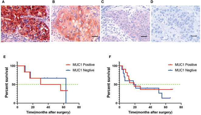
Figure 2.
MUC1 expression in human oral tongue squamous cell cancer. (A) Strong positive staining of MUC1. Positive staining of MUC1 was mainly located on the membrane and in the cytoplasm of cancer cells. (B) Moderate positive staining of MUC1. (C) Weak positive staining of MUC1. (D) Negative control. Scale bar: 30 µm. There was no significant difference of overall survival between MUC1 positive and MUC1 negative OTSCC patients either in stage III (P=0.07) (E) or in stage IV (P=0.32) (F). Kaplan–Meier method and log-rank test.

