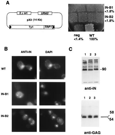
FIG. 4.
Two sites required for nuclear localization and transposition. (A) (Left) The pX3 GAL-Ty1-TRP1 plasmid (58) was used as the parent for making the IN B1 and B2 mutants (residues underlined in amino acid sequence of Fig. 1 were replaced with AAGSAA). Cells containing these pX3 derivatives were assayed for Ty1-TRP1 transposition. (Right) Transposition results for pX3 (wild-type IN [WT]) and mutants B1, B2, and RT (the negative control [neg]) are shown. Percentages of wild-type transposition frequency are indicated. (B) Indirect immunofluorescence. Anti-IN (MAb 8B11) panels are shown on the left (stained with FITC); DAPI staining is shown on the right. (C) (Top) Immunoblot with anti-IN (MAb 8B11) of whole-cell extracts prepared from wild-type (lane 1), IN B1 (lane 2), and IN B2 (lane 3) strains. The apparent molecular mass of fully processed IN is indicated on the right in kilodaltons. (Bottom) Immunoblot with R1-F (anti-Gag antibody). The apparent molecular masses of unprocessed and processed Gag proteins are indicated on the right in kilodaltons. Lanes are as described for the top panel.

