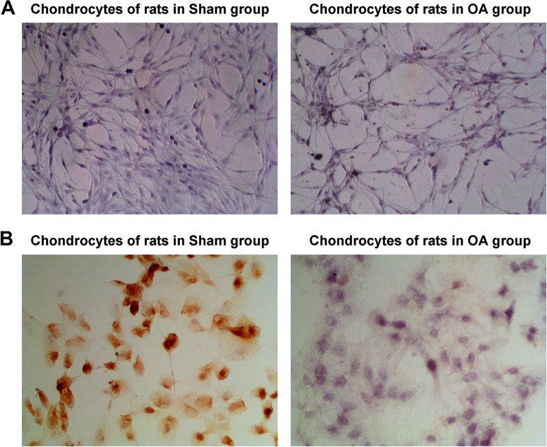
Fig. 3.
Isolation and identification of chondrocytes. A Toluidine blue staining identified chondrocytes. The chondrocytes isolated from rats in Sham group and OA group were blue and purple; while, the chondrocytes in OA group were reduced in number, sparsely arranged and with light staining; B Type II collagen staining observed the morphology of chondrocytes. COL2 was positive in the chondrocytes of rats in the Sham group, with brown particles visible in the cytoplasm, and the nucleus was basically unstained; COL2 staining was weak and positive in the chondrocytes of rats in the Model group, with a few light yellow particles in the cytoplasm, and the nucleus was basically unstained

