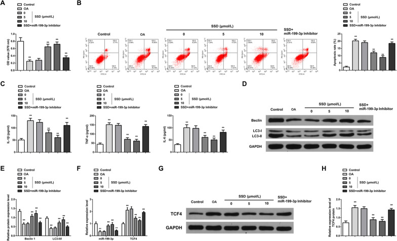
Fig. 4.
SSD relieves OA chondrocyte inflammation and controls autophagy via elevating miR-199-3p. A MTT analyzed cell proliferation. OA chondrocyte proliferation decreased; while, SSD treatment promoted cell proliferation. Downregulation of miR-199-3p attenuated the promoting effect of SSD on chondrocyte proliferation; B Flow cytometry measured cell apoptosis. OA chondrocyte apoptosis increased; while, SSD treatment inhibited apoptosis. Downregulation of miR-199-3p weakened the inhibitory effect of SSD on chondrocyte apoptosis; C ELISA measured content of pro-inflammatory cytokines IL-1β, TNF-α, IL-6 in the cell supernatant. The contents of IL-1β, TNF-α and IL-6 in the supernatant of OA chondrocytes were increased; while, the contents of IL-1β, TNF-α and IL-6 were decreased by SSD treatment. Downregulation of miR-199-3p weakened the effect of SSD; D–E Western blot detected autophagy-correlated Beclin1 and LC3-II/LC3-I ratio. Beclin1 and LC3-II/LC3-I ratio decreased in OA chondrocytes; while, SSD treatment increased Beclin1 and LC3-II/LC3-I ratio. Downregulation of miR-199-3p weakened the effect of SSD; F RT-qPCR evaluated miR-199-3p and TCF4 mRNA expression. The expression of miR-199-3p was downregulated and the expression of TCF4 mRNA was upregulated in OA chondrocytes. SSD treatment could promote the expression of miR-199-3p and inhibit the expression of TCF4 mRNA. Downregulation of miR-199-3p weakened the effect of SSD; G–H Western blot detected TCF4 protein expression. The expression of TCF4 protein was upregulated in OA chondrocytes; while, SSD treatment inhibited the expression of TCF4 protein. Downregulation of miR-199-3p weakened the effect of SSD. * P < 0.05, ** P < 0.01. N = 3

