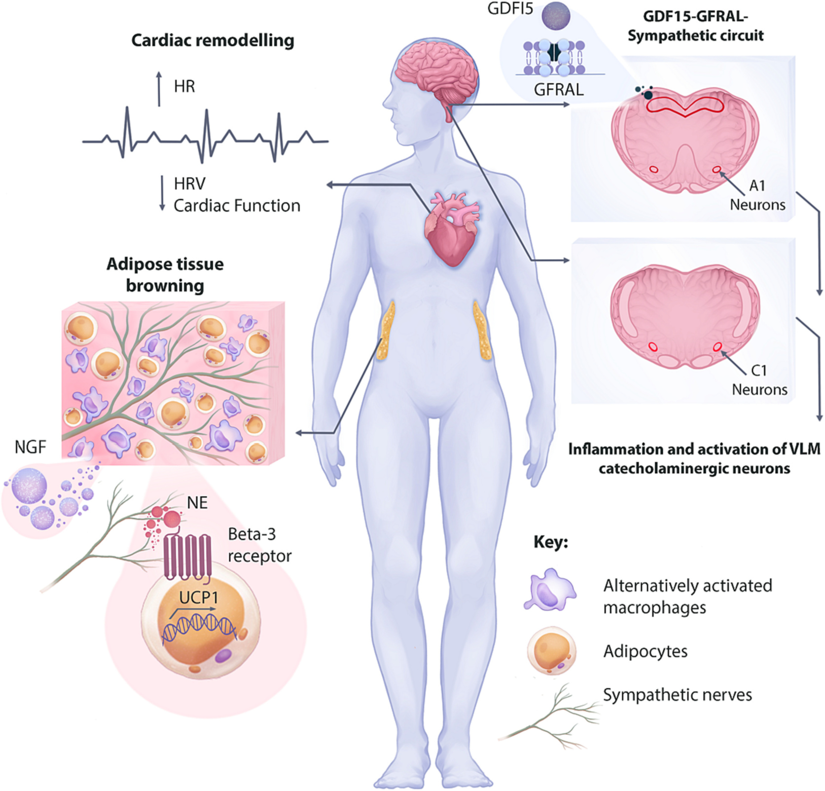
Fig. 5.

Summary of findings and potential mechanisms of increased sympathetic signaling and tissue remodeling during cancer-associated cachexia. An increased adrenergic transmission in the heart increases heart rate while decreasing heart rate variability and regulating maladaptive cardiac remodeling. Although the exact mechanism of enhanced adrenergic transmission in the heart is unknown, inflammation and activating of VLM catecholaminergic neurons are reported in animal models. Additionally, the GDF15-GFRAL-Sympathetic circuit in involved in increasing skeletal muscle adrenergic transmission, leading to increased energy expenditure. Increased nerve density in the white adipose tissue (WAT) is regulated by the nerve growth factor (NGF) secreted from alternatively activated macrophages. The increased nerve density and adrenergic signaling on beta-3 adrenergic receptor regulates WAT browning and increased energy expenditure in some animal models of cancer cachexia.
