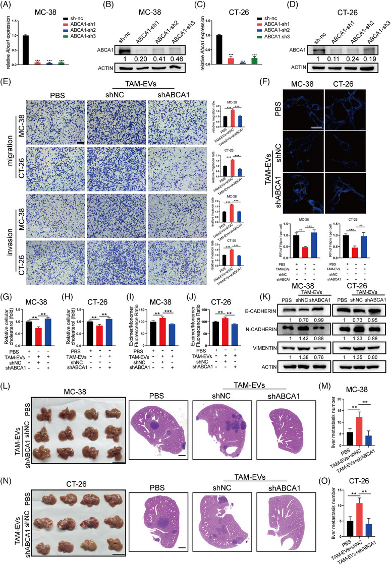
FIGURE 4.

TAM‐EVs regulate cholesterol metabolism, membrane fluidity and metastatic ability via ABCA1 in CRC cells. (A–D) qRT‒PCR (A and C) and Western blot (B and D) analyses were conducted to determine the knockdown efficiency of the shRNA targeting ABCA1 in MC‐38 and CT‐26 cells. (E) Transwell assays were used to determine the effect of decreased ABCA1 expression on CRC cells; the quantification of the migrated and invaded cells is shown on the right. Scale bar: 200 μm. (F) Membrane cholesterol was evaluated by filipin staining in CRC cells. Scale bar: 20 μm. The quantification of the mean fluorescence intensity (MFI) is shown on the right. (G and H) The total cholesterol content was measured in CRC cells treated with TAM‐EVs and transduced with or without shABCA1. (I and J) Membrane fluidity was evaluated in control and shABCA1‐transfected CRC cells treated with TAM‐EVs. (L–O) Representative morphological and HE staining images of livers from a murine ectopic hepatic metastasis model established by intrasplenic injection of CRC cells transfected with empty vector or the shABCA1 plasmid and subsequent treatment with PBS or TAM‐EVs. The quantification of metastatic nodules in the liver is shown on the right. Scale bar: 2 cm. The data are presented as the means ± SDs. *p < .05, **p < .01, ***p < .001.
