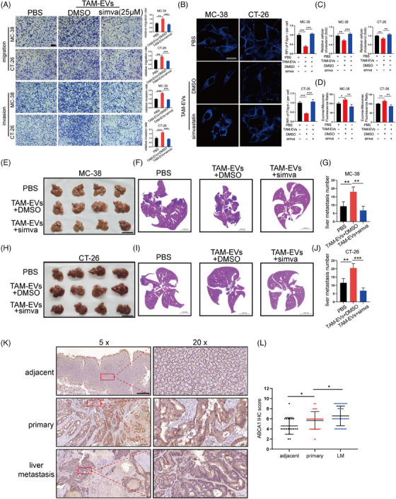
FIGURE 7.

Simvastatin attenuated the prometastatic effect of TAM‐EVs via inhibition of ABCA1. (A) Representative images of migration and invasion assays of CRC cells treated with TAM‐EVs with or without simvastatin, accompanied by the quantification of migrated and invaded cells. Scale bar: 200 μm. (B) Filipin staining was used to evaluate membrane cholesterol in CRC cells treated with TAM‐EVs with or without simvastatin. Scale bar: 20 μm. The quantification of the mean fluorescence intensity (MFI) is shown on the right. (C) The total cholesterol content was measured in CRC cells treated with TAM‐EVs with or without simvastatin. (D) Membrane fluidity was evaluated in CRC cells treated with TAM‐EVs with or without simvastatin. (E–J) Representative morphological (E and H) and HE staining images (F and I) of livers from a murine ectopic hepatic metastasis model established by intrasplenic injection of CRC cells treated with or without TAM‐EVs followed by intragastric administration of simvastatin every other day. The quantification of liver metastatic nodules is shown on the right (G and J). Scale bar: 5 cm. (K and L) IHC analysis of ABCA1 expression in adjacent normal tissues, primary CRC tissues and liver metastatic specimens from CRC patients with liver metastasis. Scale bar: 500 μm (K). The quantification of the IHC score for ABCA1 is shown on the right (L). The data are presented as the means ± SDs. *p < .05, **p < .01, ***p < .001.
