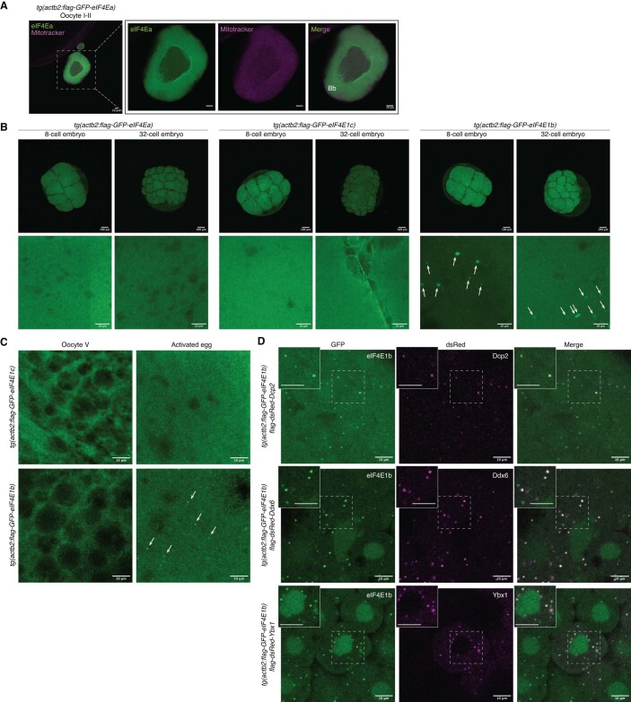
Figure EV3. eIF4E1b localizes to P-bodies in the embryo.
(A) eIF4Ea localizes to the cytosol of fixed early zebrafish oocytes. Mitochondria present in the Balbiani body (Bb) are stained with Mitotracker. Scale bars = 10 μm. (B) Confocal microscopy images of fixed transgenic embryos expressing 3xflag-sfGFP-tagged eIF4Ea, eIF4E1b and eIF4E1c (scale bars: top = 100 μm; bottom = 10 μm). eIF4E1b foci are indicated by white arrows. (C) Confocal microscopy images of squeezed (oocyte V) and activated eggs from transgenic lines expressing 3xflag-sfGFP-tagged eIF4E1c (top) and eIF4E1b (bottom). eIF4E1b foci are indicated by white arrows. (D) Colocalization experiments in zebrafish embryos expressing 3xflag-sfGFP-eIF4E1b transiently expressing P-body markers. mRNAs containing the coding sequences of dsRed-tagged P-body components (Dcp2, Ddx6 and Ybx1) were injected into 1-cell embryos. Images were taken at 3 h post fertilization. Regions enclosed by dashed boxes are shown at higher magnification on the top left (scale bars = 10 μm).

