
Figure 1.
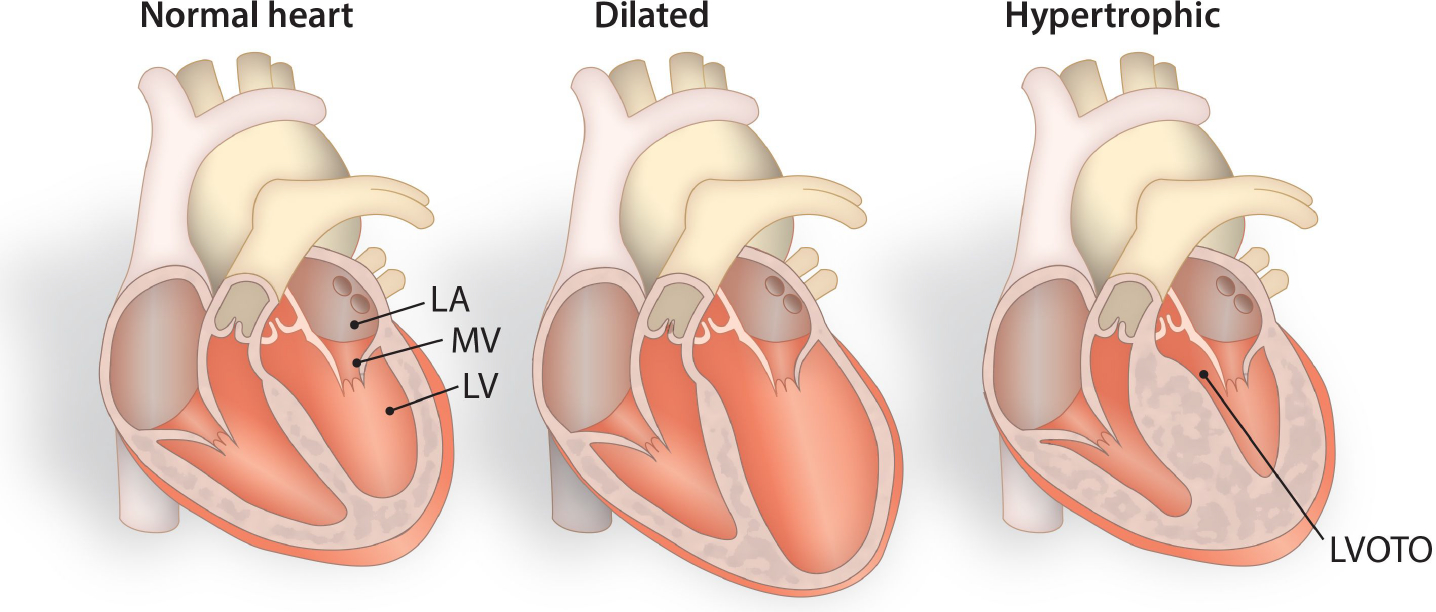
Schematic diagram differentiating normal hearts from dilated and hypertrophied hearts. Cardiomyopathies occur due to genetic variations, resulting in distinct physiological and/or pathophysiological consequences. In terms of clinical manifestations, cardiomyocytes within hypertrophied hearts become enlarged and demonstrate cardiac dysfunction due to increased left ventricular wall thickness, diminished left ventricular cavity size, and altered blood flow rates. LA-Left Atrium, MV-Mitral Valve, LV-Left Ventricle, LVOTO-Left Ventricular Outflow Tract Obstruction.