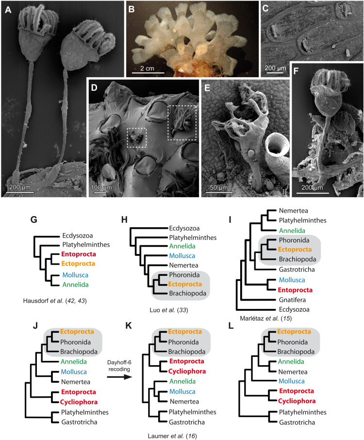
Fig. 1. General view and phylogenetic position of Entoprocta and Ectoprocta proposed in previous studies.
(A) Part of B. gracilis colony, scanning electron microscopy (SEM) image. (B) General view of T. membranaceotruncata colony and (C) SEM image of T. membranaceotruncata autozooids. (D) SEM image of L. nordgardii individuals on the surface of a bryozoan colony. (E) SEM image of L. nordgardii on the surface of algae with Folliculina sp. ciliate next to it. (F) SEM image of the colony part in P. cernua. (G) A scheme proposed by Hausdorf et al. (42, 43). (H) Affinity of Ectoprocta to Phoronida by Luo et al. (33). (I) Separate placement of the taxa by Marlétaz et al. (15). (J) Separate positions of Entoprocta + Cycliophora and Ectoprocta by Laumer et al. (16). (K) Ectoprocta and Entoprocta in one clade after Dayhoff-6 transformation. (L) Third version of lophotrochozoan topology by Laumer et al. (16).

