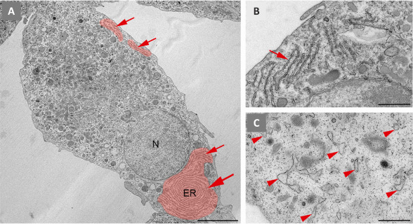
Figure 1.

Ultrastructural features of the endoplasmic reticulum in primary microglia in culture.
We isolated microglia from cerebral cortex of neonatal mice. Subsequently, they were cultured in vitro and processed for transmission electron microscopy (TEM) visualization. (A) Ultrathin section showing a euchromatic nucleus (N) and a cytoplasm heavily filled with components of the endomembrane system, including dispersed long stretches (arrows) of rough endoplasmic reticulum (ER), that can adopt a characteristic fingerprint shape (big arrowhead). The ER was pseudocoloured in red. (B) High magnification of unstimulated microglia showing narrow long ER stretches (arrow). (C) High magnification of microglia stimulated with 100 ng/mL lipopolysaccharide for 24 hours to induce an inflammatory phenotype before TEM processing. The cell contains dilated and dispersed ER (arrowheads). Sourced from the authors' laboratory (unpublished data). Scale bars: 5 µm in A; 1 µm in B and C.
