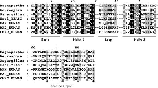
Fig. 2.
Alignment of Myc, Max, and Mad together with four putative fungal Myc sequences, visualized by genedoc (www.psc.edu/biomed/genedoc), with conserved sites indicated by differential shading. Two gaps separate the various labeled functional domains. Heptad leucine repeats in the zipper are labeled with a bold plus (+) sign.

