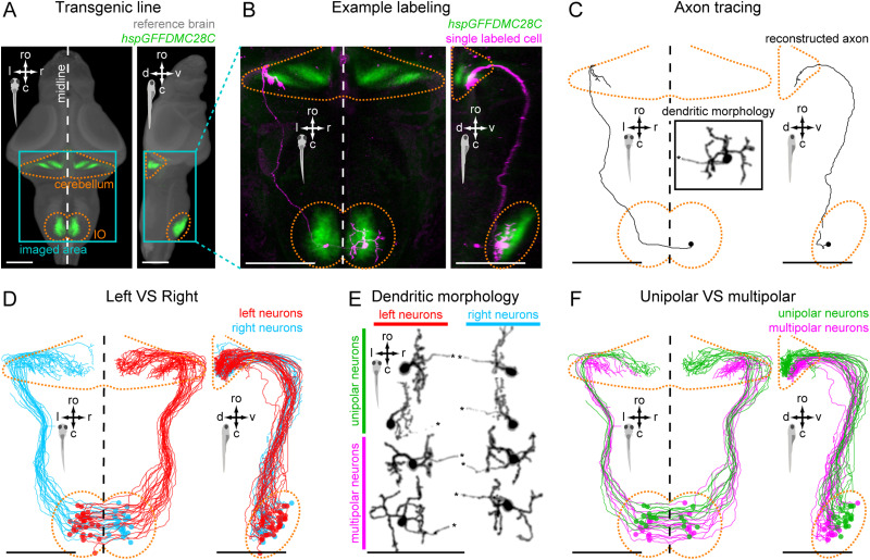
Figure 1.
IO neurons can be divided into distinct morpho-anatomical types. A, Dorsal and lateral views of the average expression of the hspGFFDMC28C line used in this study (green, N = 39 fish; line from Takeuchi et al., 2015) registered to a common reference larval zebrafish brain (gray), showing strong signal in the IO and in the CFs in the cerebellum. In this and subsequent panels: ro, rostral direction; l, left; r, right; c, caudal; d, dorsal; v, ventral; scale bars, 100 µm; vertical dashed lines indicate the midline of the brain. Teal rectangle outlines the area shown in B, C, D, and F. B, Example of a single labeled IO neuron (magenta). C, Axon reconstruction of that neuron. The inset shows its dendritic morphology, and the asterisk indicates its axon. D, Axon reconstruction of all labeled IO neurons (N = 53 neurons from 39 larvae) color coded by soma location (red, left IO, teal, right IO), showing that IO neurons project contralaterally. E, Examples of IO neurons that were divided into two morphological classes: unipolar neurons (green) that have a single dendritic tree arborized along the midline, and multipolar neurons (magenta) that have bi- or tri-polar dendritic trees. Asterisks indicate axons. For the complete dataset (N = 16 unipolar, 19 multipolar and 18 ambiguous neurons) see Figure 1-1. F, Axon reconstruction of all unipolar (green) and multipolar neurons (magenta), showing that the morphological type of a neuron is predictive of its projection pattern and its location within the IO.

