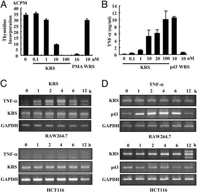
Fig. 3.
The effect of KRS on macrophage proliferation. (A) The proliferation of RAW264.7 cells was monitored by the incorporation of tritium-labeled thymidine at different KRS concentrations. Phorbol 12-myristate 13-acetate (PMA) and WRS (7) were used as positive and negative controls, respectively. (B) The effect of KRS on the secretion of TNF-α from RAW264.7 cells. p43 (32) and WRS were used as positive and negative controls, respectively. (C) RAW264.7 and HCT116 cells were treated with 10 nM KRS for the indicated times, and the transcript levels for TNF-α and KRS were determined by RT-PCR as described in Materials and Methods.(D) The two cells were treated with 10 ng/ml TNF-α, and the expression of KRS and p43 was determined by RT-PCR. GAPDH was used as a control.

