Figure 1.

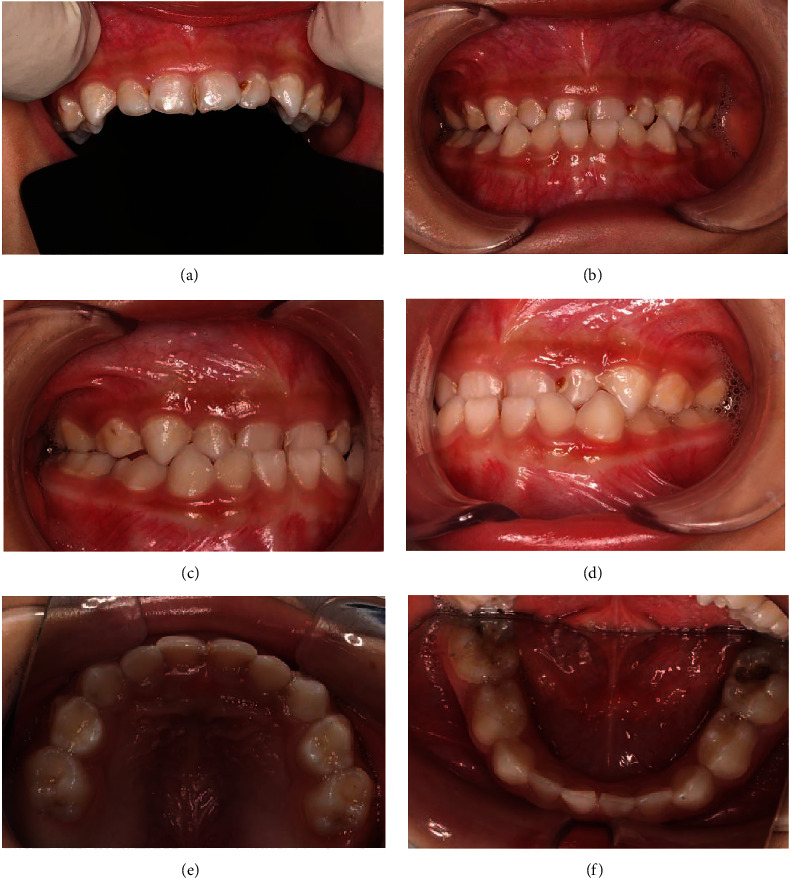
Intraoral photos of the patient before treatment: maxillary anterior teeth (a), frontal occlusion (b), right lateral occlusion (c), left lateral occlusion (d), upper occlusion (e), and lower occlusion (f).

Intraoral photos of the patient before treatment: maxillary anterior teeth (a), frontal occlusion (b), right lateral occlusion (c), left lateral occlusion (d), upper occlusion (e), and lower occlusion (f).