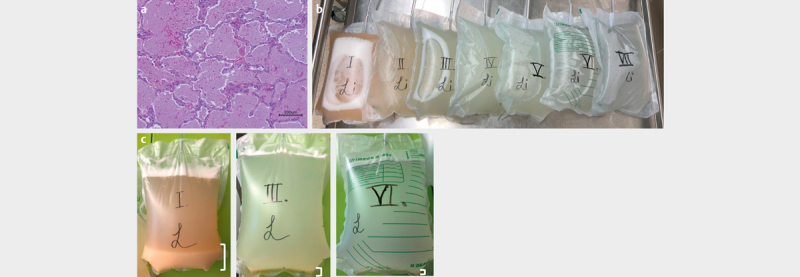
Fig. 1.

Whole lung lavage of hereditary pulmonary alveolar proteinosis (A) Tissue section of the distal lung compartment to assess the proteinosis phenotype within the alveoli. Tissue sections are typically stained by the Periodic acid-Shiff (PAS) method to stain for muco-like substances in the lung such as glycoproteins, glycolipids and others. Picture shows an extensive proteinosis by alveoli filed with surfactant material. (B) Photo showing the recovery of sequential lung lavage (WLL) batches in a child with herPAP. Each lavage recovery is numbered from the left to the right (I to VII), showing the overall improvement demonstrated by a decrease turbidity. (C) Highlighting examples from WLL recoveries demonstrating the decrease in muco-like substances which is highlighted by the white brackets. Three pictures are shown from first (I), third (III) and 6th (VI) lavage.
