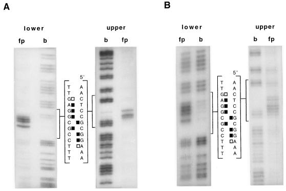
FIG. 4.
Purine missing-base interference footprints of GST-NirA(1–125) fusion protein (A) and NirA(1–125) peptide (B) on site 2 of the niiA-niaD intergenic region. “upper” and “lower” refer to strands as in Fig. 2. fp, free probe; b, bound probe. Only the sequence of the limited region bracketing the NirA binding sequence is shown. Solid squares, strongly interfering purines; open squares, partially interfering purines. The probe for site 2 is as described above. Footprint experiments have been carried out at least twice with identical results.

