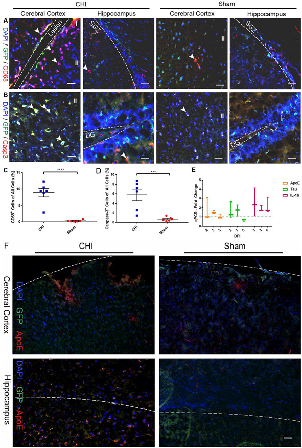
Fig. 2. CHI induces substantial inflammation and cell death in the cerebral cortex and hippocampus.
Injury-induced inflammation and cell death in the cerebral cortex and hippocampus were determined by immunostaining and qPCR. Cells expressing macrophage marker CD68 (A) and cell death marker Caspase-3 (Casp3) (B) were detected in coronal sections at 2 days post injury (DPI). Quantification shows a significant increase in the number of CD68+ macrophages (p < .0001) (C) and Caspase-3+ cells (p < .0005) (D) in the cerebral cortex of injured animals as compared to the sham animals. Mean ± SEM, n = 6. (E) qPCR analysis shows fold-change increase in markers of TBI (ApoE, Tau) and inflammation (IL-1b) at 2, 3, and 5 DPI compared to sham animals. (F) Representative photomicrographs of TBI marker ApoE staining. Scale bars = 30 μm.

