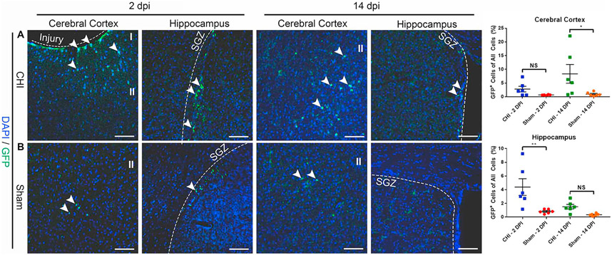
Fig. 3. CHI increases the number of Notch1CR2-GFP+ cells in the cerebral cortex and hippocampus.
Injury-induced GFP+ cells in the cerebral cortex and hippocampus were determined by immunostaining in a mouse model of closed head injury (CHI). GFP+ cells in the injured cerebral cortex and hippocampus (SGZ) were detected in coronal sections of the brain at 2 DPI and 14 DPI. An increased number of GFP+ cells was observed in the injured hippocampus at 2 DPI (p < .01) and in injured cerebral cortex at 14 DPI (p < .05) (A) as compared to sham mice (B). Scale bar = 50 μm, Mean ± SEM, n = 6.

