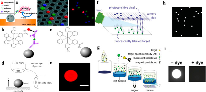
Figure 9.
(a) Schematic illustration of a sandwich immunoassay, wherein a bead-based platform was constructed to achieve the ECL detection of multiple antigens simultaneously in a microarray format. Reprinted from ref (141) from American Chemical Society, copyright 2009. (b) Schematic illustration of a sandwich bead-based immunoassay with an ECL readout. (c) Schematic showing the functionalization of a PS bead with an ECL label. (d) Schematic of the two different microscope objective configurations (i.e., top-view and side-view) utilized for imaging the labeled bead. (e) PL image (top view) of a ruthenium (homogeneously distributed) labeled PS bead via a sandwich immunoassay format. The scale bar was 10 μm. Reprinted from ref (142) with permission from Elsevier, copyright 2020. (f) Schematic illustration of the detection/imaging of individual fluorescent beads (bound with molecular targets) on the camera’s chip by capturing one or a small group of pixels without the requirement of magnified microscope. (g) After the formation of the immunocomplex, the magnetic beads were drawn toward the microwell bottom (through dye cushion) by applying a magnet and deposited on the imaging surface. Only fluorescent beads in proximity of the surface were excited due to the excitation light absorption (deep into the well). (h) Digital camera recording of fluorescent beads as bright pixels. (i) Images of wells displaying the comparison with and without dye for revealing the effectiveness of the dye-cushion layer. Reprinted from ref (95) with permission from the Springer Nature under a Creative Commons Attribution 4.0 International License.

