Fig. 1.
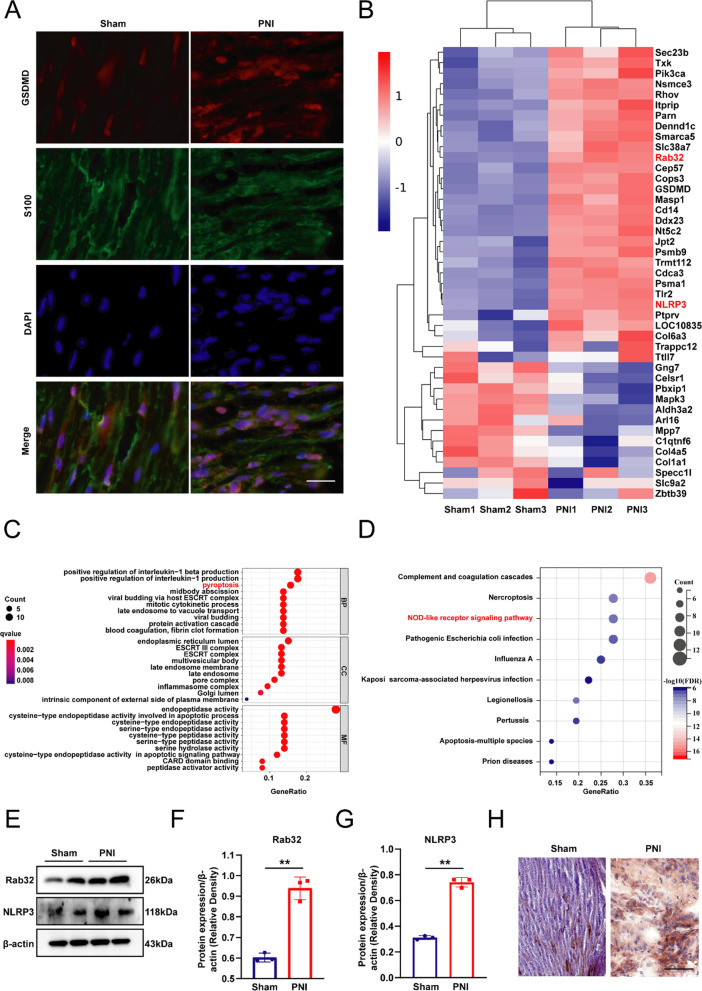
PNI induced pyroptosis and upregulated Rab32 expression. A Immunofluorescence staining for S100 (green) and GSDMD (red) in nerve tissues. Nuclei were labeled using DAPI (blue). Scale bar = 20 µm. B Heat map summarizing the DEGs related to Rab32 and pyroptosis-related gene. C The GO biological process enrichment analysis of DEGs. D The KEGG biological process enrichment analysis of DEGs. E–G Protein expression levels and quantitative results of Rab32 in Sham and PNI groups. (H) Immunohistochemical staining of Rab32 in peripheral nerve tissue. **P < 0.01. Scale bar = 50 µm

