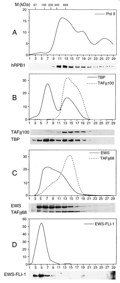
FIG. 6.
Sedimentation of RD-ES cell NE through a 20 to 40% glycerol gradient indicates that the endogenous EWS–FLI-1 fusion protein is present in low-molecular-mass ranges. The relative sedimentations of the largest subunit of Pol II (A), TAFII100 and TBP (B), EWS and TAFII68 together (C), and EWS–FLI-1 (D) were determined by Western blotting using antibodies raised against either the CTD of the largest subunit of Pol II (A), TAFII100 and TBP (B), or EWS and TAFII68 (C). To better visualize EWS–FLI-1 that is only weakly detected in panel C by the EWS antibody, in panel D the anti-FLI-1 antibody was used. In each panel, the upper part shows a quantification of the Western blot. Values represent the percentage of a given protein present in each fraction compared to the total amount of this protein loaded on the glycerol gradient. Positions of markers (M) of known molecular mass standards are indicated at the top of panel A.

