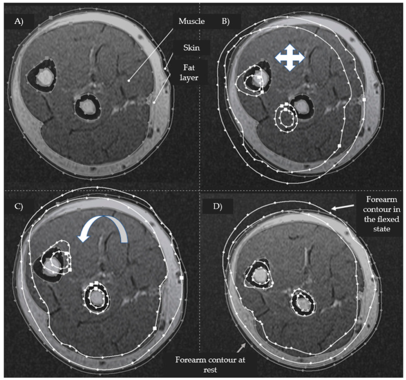
Figure 3.
An example of contouring the skin-fat, muscle, and bone layer according to an MRI image and applying the corresponding contours (gray contour—the contour of the forearm at rest; white contour—the contour of the forearm in a state of flexion) after combining the outlines of the ulna and radius bones ((A)—contouring the layers using the “Spline” function; (B)—superimposition of the corresponding sections; (C)—alignment along the outlines of the radius; and (D)—alignment along the outlines of the ulna).

