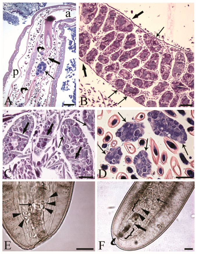
Figure 1.
Inseminated female acanthocephalans of Centrorhynchus globocaudatus. (A) Longitudinal section through fully mature female body; free ovarian ball (arrow) encircled by numerous eggs. Note extension of the ligament sac (curved arrows) from the base of the receptacle in anterior part of body (a) to the posterior end (p) of female body and point of its disruption (thick arrow). Giemsa staining; scale bar, 200 µm. (B) Immature female; numerous ovarian balls are still within the lining of the ligament sac (thick arrows) and some are loosely attached to it (arrows). Alcian Blue/PAS staining; scale bar, 100 µm. (C) Free ovarian balls in a fully mature female trunk; some eggs including acanthors (arrows) are in the periphery and few (thick arrows) in the inner region of the ovarian balls. Alcian Blue/PAS staining; scale bar, 50 µm. (D) Free ovarian balls (arrows) are surrounded by numerous shelled developmental stages. Giemsa staining; scale bar, 50 µm. (E) Mounted female C. globocaudatus; transition of uterine bell into tube-like uterus (arrow). The organ is surrounded by several shelled acanthors (arrowheads). Scale bar, 100 µm. (F) Posterior end of mounted female. A shelled acanthor (thin arrow) enters the inner opening of uterine bell; two acanthors (arrowheads) inside the uterine bell. Uterus (thick arrow) and genital opening (curved arrow) are visible. All the reproductive organs are surrounded by their envelopes inside the genital sheath (brackets); scale bar, 100 µm.

