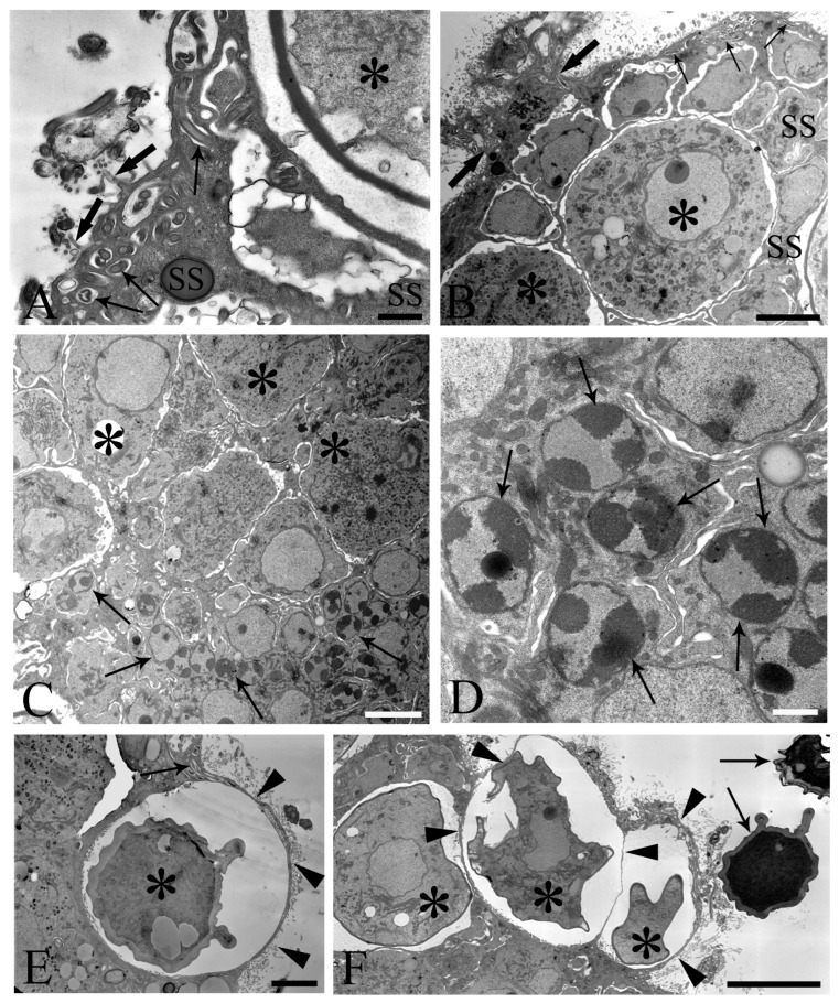
Figure 4.
Electron microscope images of ovarian balls in fecundated female Centrorhynchus globocaudatus. (A) Spermatozoa flagella (arrows) are embedded in the support syncytium (SS); penetration of spermatozoa into ovarian ball via microvillous surface (thick arrows); inseminated mature oocyte (asterisk) with completed shell; scale bar, 0.8 µm. (B) Penetration of microvillous OB surface by sperm flagella (thick arrows); flagellum (arrows) embedment in the SS is visible; two inseminated mature oocytes (asterisks) present with numerous electron-dense membrane-bound granules; scale bar, 5 µm. (C) Micrograph showing mainly the middle region of an OB with several SS nuclei (arrows) and some primary oocytes (asterisks); scale bar, 5 µm. (D) High magnification of the middle region of the OB; aspect of the multinucleate SS is evident; abundant heterochromatin laid on the inner membrane side of some of its nuclei (arrows); scale bar, 1 µm. (E) Inseminated mature oocyte (asterisk) with completed shell shortly before disruption of very thin peripheral SS envelope (arrowheads) and release from the OB. Note flagellum (arrow) embedment in SS; scale bar, 3.3 µm. (F) Periphery of an OB; some inseminated mature oocytes (asterisks) with shells in formation are peripherally enveloped by a very thin SS layer (arrowheads); after rupturing of the envelope, shelled eggs leave the organ; two free acanthors (arrows) near the OB; scale bar, 3 µm.

