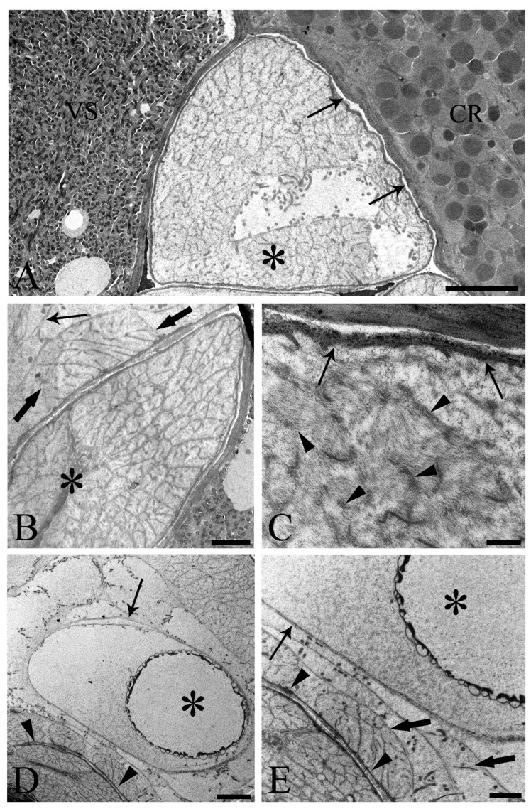
Figure 5.
Transmission electron micrographs of sections of mature male Centrorhynchus globocaudatus posterior ends. (A) Contact between Saefftigen’s pouch (asterisk), vesicula seminalis (VS), and cement reservoir (CR). Each organ has its own lining; often, there is only a narrow distance between these organs (arrows); scale bar, 5 µm. (B) Micrograph shows most of the spongy Saefftigen’s pouch (asterisk) in vicinity of the copulatory bursa (arrow); in interface region between both organs appears to be tissue of almost identical aspect to the main portion of Saefftigen’s pouch (thick arrows); scale bar, 2 µm. (C) High magnification underscores the spongy aspect of Saefftigen’s pouch, filled with some electron-dense matrix (arrowheads); pouch is delimited by its own lining (arrows); scale bar, 0.5 µm. (D) Posterior end of male with portion of the pouch (arrowheads) in proximity to the inverted bursa (arrow); opening of the bursa (asterisk); scale bar, 5 µm. (E) Higher magnification of (D): in interface region between pouch (arrowhead) and bursa (arrow), occurrence of genital sheath (thick arrows) is evident; opening of the bursa (asterisk); scale bar, 2 µm.

