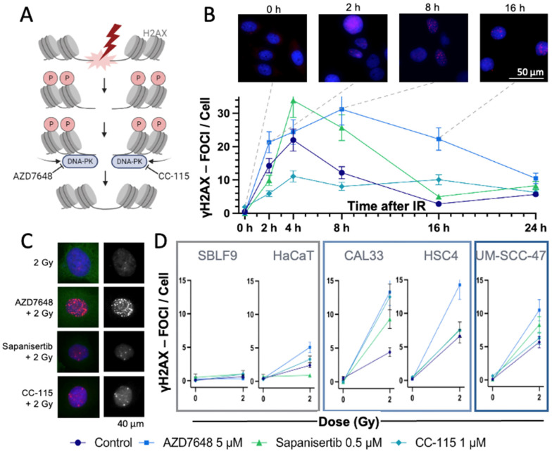
Figure 2.
Analysis of DNA DSBs using γH2AX-IS. (A) IR leads to DNA DSBs, with DNA-PK involved in repair. When DNA DSBs occur, the histone protein H2AX is γ-phosphorylated and can be visualized by IS. DNA-PK-Is like AZD7648 or CC-115 block the repair of DSBs, thereby keeping H2AX phosphorylated and detectable as γH2AX foci via IS (created with BioRender.com, accessed on 12 December 2023). (B) Mean number of γH2AX foci in the nuclei of the HNSCC UM-SCC-47 cells over a period of 0 to 24 h after IR ± KI. Time 0 was not irradiated. Representative images of AZD7646-treated UM-SCC-47 stained with DAPI (blue) and γH2AX (red)—fluorescence microscopy image. Dashed lines indicate the times of the images with γH2AX foci. Error bars represent the SD of at least three replicates. (C) Exemplary presentation of fluorescence microscopy images of HNSCC CAL33 cells after IR ± KI. Left: The γH2AX foci are red and localized in the blue nuclei. Right: Monochromatic image of γH2AX foci. (D) Mean number of γH2AX foci in different normal (SBLF9, HaCaT) and HNSCC (CAL33, HSC4, UM-SCC-47) cell lines, 24 h after IR ± KI. Error bars represent the SD of at least three replicates.

