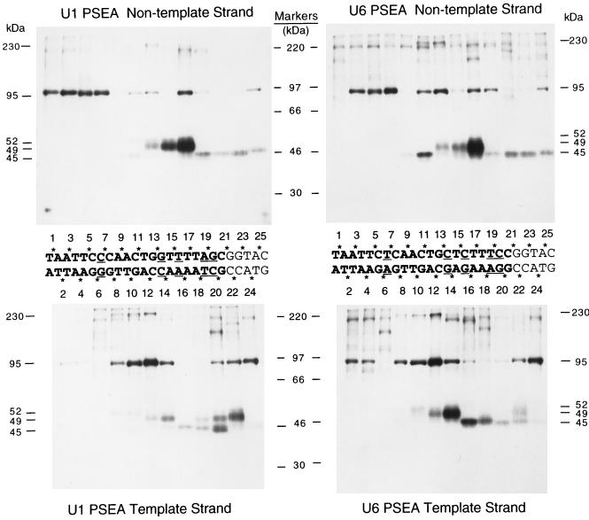
FIG. 3.
Site-specific protein-DNA photo-cross-linking reactions that scan through the PSEA sequences of Drosophila U1 and U6 genes. Fifty different radiolabeled site-specific photo-cross-linking probes were incubated in separate reactions with the HA300 fraction that contains the DmPBP activity. Following UV irradiation, polypeptides that cross-linked to the DNA were detected by SDS gel electrophoresis and autoradiography. The sequences of the U1 (left) and U6 (right) PSEAs (and five bases downstream of the PSEAs) are shown between the upper and lower panels. The five-base-pair differences between the U1 and U6 sequences are indicated by underlining. The phosphate positions on the template and nontemplate strands that were derivatized with cross-linking reagent are indicated by asterisks. The numbers below the upper panels and above the lower panels indicate the individual lanes that contain the cross-linking results from the correspondingly numbered phosphate positions. (According to our numbering system, the phosphate designated 1 is the phosphate that links the first and second nucleotides in the PSEA. This differs from the standard International Union of Pure and Applied Chemistry convention, which would require that all the phosphate positions on the nontemplate strand be increased by a value of 1.) The upper panels show the results of cross-linking to the nontemplate strand of the U1 PSEA (left) and to the nontemplate strand of the U6 PSEA (right). The lower panels show the results of cross-linking to the complementary template strands. The positions migrated by molecular weight markers are shown in the center. The molecular masses of the specifically cross-linked polypeptides, identified in this and later experiments (Fig. 4 and 5), are indicated to the far left and far right.

