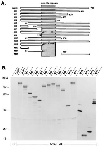
FIG. 1.
DMP1 mutants. (A) Schematic representation of wild-type DMP1 (top line) and various mutants (M1 to M15). All are deletion mutants except for M11, which contains a Glu-for-Lys substitution at codon 319 (K319E, marked with an asterisk) located within the second myb repeat. The numbers indicate the deletion boundaries, and the central region containing the three tandem myb repeats is shaded. (B) Metabolically labeled wild-type and mutant DMP1 proteins recovered from baculovirus vector-infected Sf9 cells and separated on denaturing polyacrylamide gels. The first lane on the left indicates the results of precipitation of wild-type DMP1 with an irrelevant control monoclonal antibody (designated C), as indicated at the bottom of the panel. All other lysates were precipitated with monoclonal antibody M2 directed to the Flag epitope at the DMP1 N terminus. The mobilities of markers of known molecular mass are indicated to the left of the panel.

