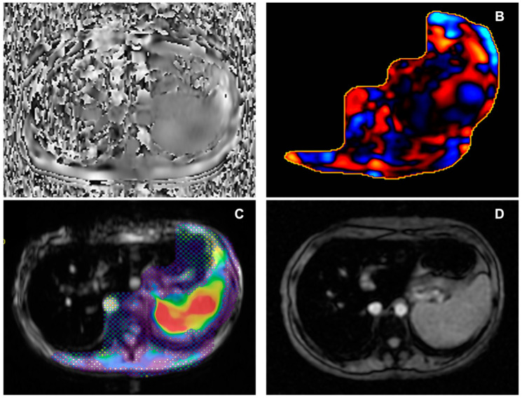
Figure 4.
Male, 38 years old, with severe iron overload. (A) Axial phase image showing the absence of wave propagation through the liver. (B) Non-diagnostic wave image. (C) Elastogram reconstruction with the interposition of the 95% confidence map not covering the liver area. (D) Axial Starmap T2* breath-hold sequence, confirming iron overload. In the case of severe iron overload, an option might be to use SE echoplanar sequences instead of GRE sequences, with the latter being more prone to non-diagnostic MRE, even if an univocal cut-off of severe iron overload of MRE failure has not been established yet [37,38]. As observed by a retrospective study by Meng Y. et al. [39] on 1377 consecutive MRE examinations, MRE often had a low failure rate (5.6%), with the majority of failure cases due to inadequate signal-to-noise ratio related to iron overload (3%) and the remaining cases caused by execution errors or respiratory artifacts. A retrospective study by Wagner M. et al. [40] confirmed the higher rate of technical failure associated with liver iron overload with p < 0.001. Also, the presence of bowel interposition between the liver and abdominal wall, motion artifacts, or interfering paramagnetic materials are considered as causes of low-quality elastograms. Despite the lack of published data on the subject from our centre, the failure rate aligns with literature data after the initial eight weeks of training.

