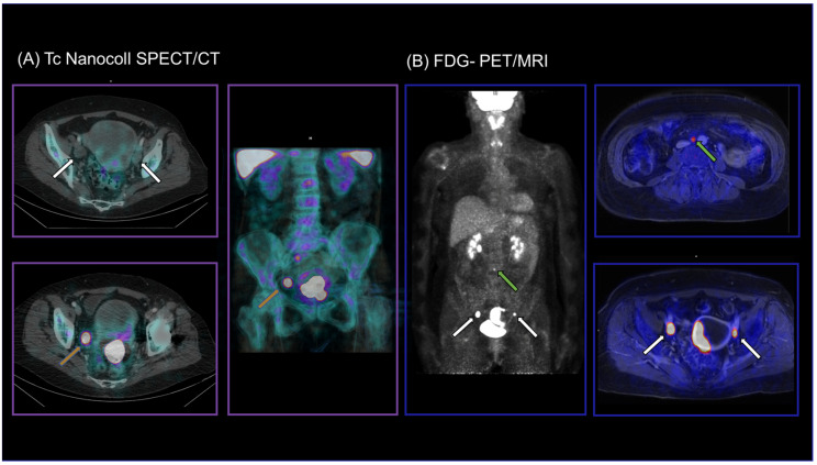
Figure 2.
SLN-SPET/CT (A) and FDG-PET/MRI (B) images of a 65-year-old patient with initial diagnosis of endometrial cancer. LN mapping was incomplete in SPECT/CT with unilateral tracer accumulation on the right hemipelvis only (orange arrows). FDG-PET/MRI detected bilateral iliac external LNM (white arrows) as well as para-aortic LNM (green arrow), which were removed and confirmed histologically (pT3a, pN1, G3).

