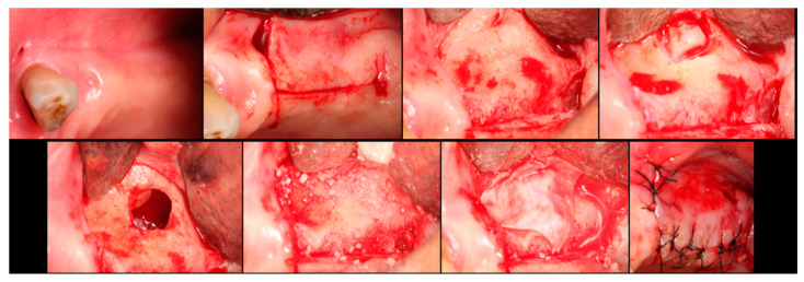
Figure 3.
Step-by-step surgical demonstration of maxillary sinus elevation with xenograft. In the first row, left to right: incision in the fourth quadrant, flap detachment, and lateral osteotomy. In the second row, left to right: accessing the sinus, filling with xenograft, collagen membrane positioning, and sutures.

