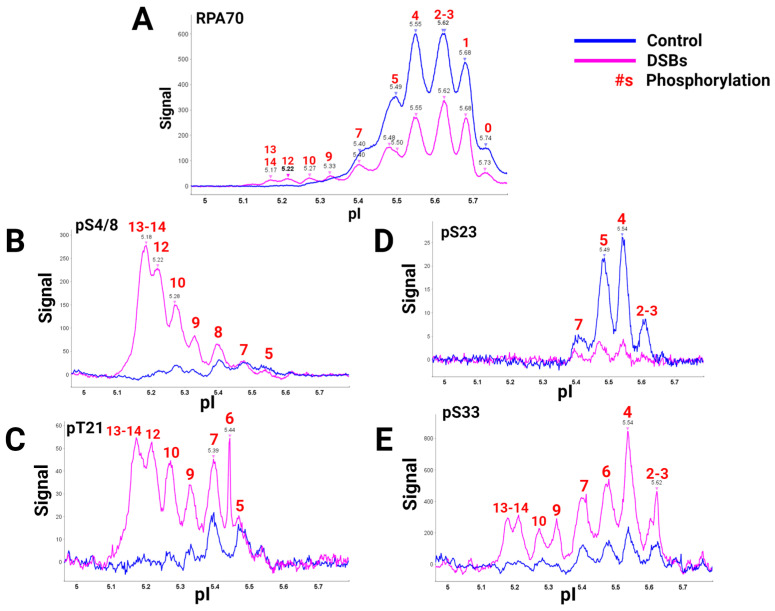
Figure 2.
RPA isoforms from cells synced in the G2 phase either with DNA damage (blue) or without DNA damage (pink), separated with isoelectric focusing using a pH gradient from 5-6 and probed with phospho-specific antibodies. RPA heterotrimer probed with (A) anti-RPA70-CT, (B) phosphoRPA32(S4/8), (C) phosphoRPA32(T21), (D) phosphoRPA32(S23), and (E) phosphoRPA32(S33) antibodies. The red number above the corresponding peaks indicates the number of phosphorylations on each isoform. Figure adapted from [40].

