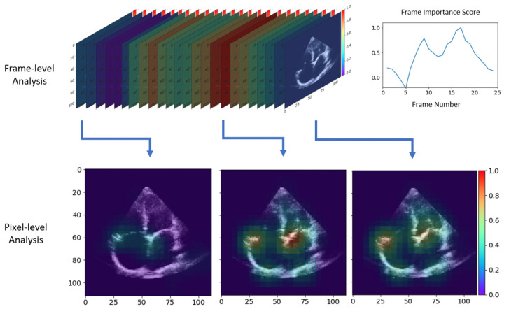
Figure 4.
Heatmaps of potential echocardiographic features of atrial remodeling reflective of atrial fibrillation identified by the deep neural networks. Spectrum ranges from red being of high importance to violet being of least importance on analysis of cines in atrial fibrillation compared to sinus rhythm. Top left is a frame-by-frame analysis with a summary of frame importance on the top right. Bottom images are pixel analysis of select frames of high importance, showing attentiveness of the deep learning model to differences in atrial structure and function with a high focus on irregularities with the opening and closure of atrioventricular valves.

