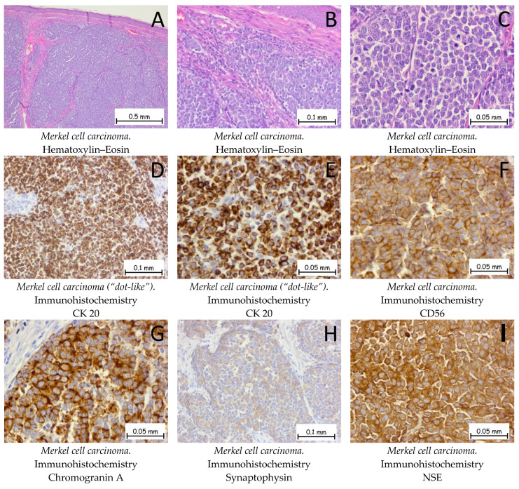
Figure 11.
Merkel cell carcinoma. (A,B). The tumor formation exhibits a solid architecture characterized by plaques and trabeculae of monomorphic cells, small to medium in size. The tumor cells do not show epidermotropism, without an ulceration of the epidermis. Between the tumoral plaques, there is a moderate chronic inflammatory infiltrate. (C). At a higher magnification, the tumoral cells have a hyperchromatic, centrally located nucleus with granular and dispersed chromatin, along with an elevated mitotic index. Tumor cells are positive for anti-CK-20 antibodies (D,E), anti-CD56 antibodies (F), anti-Chromogranin A antibodies (G), anti-Synaptophysin antibodies (H), and anti-NSE antibodies (I).

