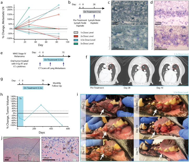
Figure 3. Case studies of patients demonstrating abscopal immune responses.
(a) Percent change in volume of regional lymph node metastasis relative to pre-treatment volume, as determined by CT measurement. (b) Fine needle aspirates were collected from the lymph node of a patient in the 1x cohort before treatment and after 2 intratumoral cytokine doses. (c) Pretreatment aspirate shows diffuse infiltration of melanocytes. (d) Lymph node disease is decreased after 2 cytokine treatments, with a marked increase in polymorphonuclear immune cells. (e) CT images from a stage IV patient in the 3.3x treatment group were collected tracking the progression of a lung metastasis after local treatment of oral melanoma. (f) CT images suggest pseudoprogression of a lung metastasis after early cytokine doses, with later regression after additional cytokine doses. (g-i) A patient in the 3.3x dosing group received a full course of treatment and had routine follow-up visits to monitor tumor progression. Tumor measurements (h) and images (i) were taken at the indicated time points, demonstrating a significantly delayed treatment response. (j) Hematoxylin and eosin staining on this tumor showed an absence of tumor cells with only scattered melanophages observed at day 529. Scale bars: 100um.

