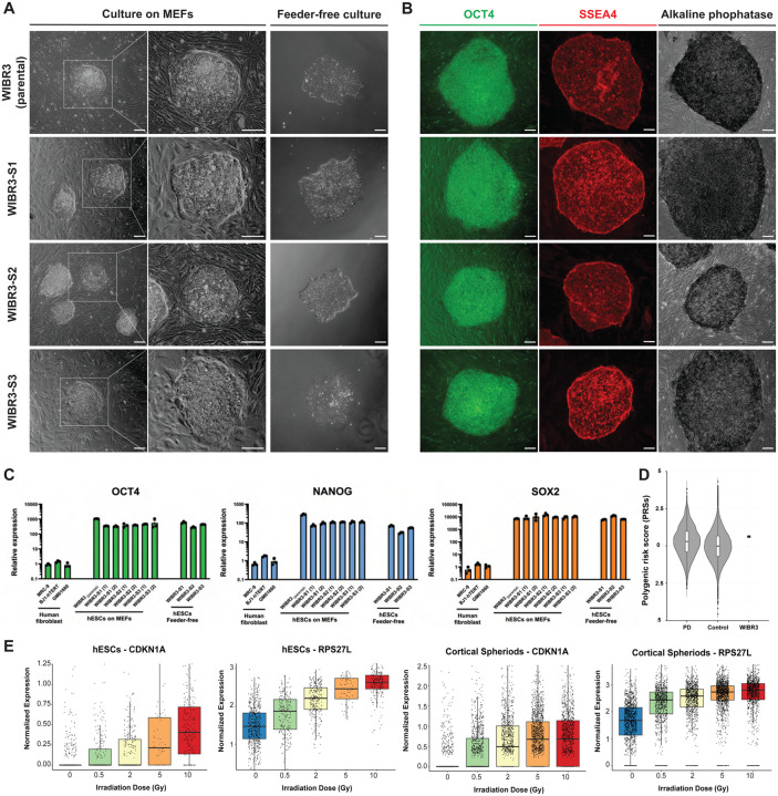
Figure 1. WIBR3 hESC cell line characterization.
(A) Phase contrast images of parental WIBR3 hESCs and subclones WIBR3-S1, WIBR3-S2 and WIBR3-S3 cultured onto MEFs and in feeder-free conditions. Scale bar 100 μm.
(B) Immunocytochemistry for pluripotency markers OCT4 (green) and SSEA4 (red) and staining for alkaline phosphatase (black) of WIBR3 (parental) hESCs and subclones WIBR3-S1, WIBR3-S2 and WIBR3-S3 cultured onto MEFs. Scale bar 100 μm.
(C) qRT-PCR analysis for the relative expression of pluripotency markers OCT4, NANOG and SOX2 in human primary fibroblasts (MRC-9, BJ1-hTERT and GM01660), WIBR3 (parental) hESCs and subclones WIBR3-S1, WIBR3-S2 and WIBR3-S3 hESCs cultured on MEFs and in feeder-free conditions. Relative expression levels were normalized to expression of these genes in primary fibroblasts. (1) and (2) indicate independent samples. (N=3; Mean +/− SEM).
(D) Polygenic risk scores (PRSs) for PD comparing WIBR3 hESCs to population-centered Z score distribution for PD PRSs in individuals with PD and the normal population from the UK Biobank.
(E) Assessment of p53 pathway activity following irradiation (0.5, 2, 5 and 10 Gy) of WIBR3 (parental) hESCs (1464 cells) and WIBR3-derived cortical spheroids (5920 cells) by scRNA-seq analysis for the expression of DNA damage response genes CDKN1A and RPS27L ( box plot showing interquartile intervals with a line at the median).

