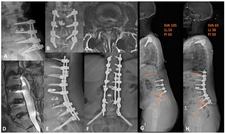
Figure 6.
(A,B) preoperative lateral and AP lumbar X-rays demonstrating the advanced degenerative changes at L2-3 with grade 2 anterolisthesis. (C) axial and (D) sagittal T2-weighted MRI showing the adjacent segment disease at L2-3 with severe central and lateral recess stenosis. (E,F) lateral and AP lumbar X-rays of the postoperative construct with apparent deformity correction. (G) preoperative and (H) postoperative whole-spine films demonstrating an improvement in sagittal balance and global lordosis. SVA = sagittal vertical axis; LL = lumbar lordosis; PI = pelvic incidence.

