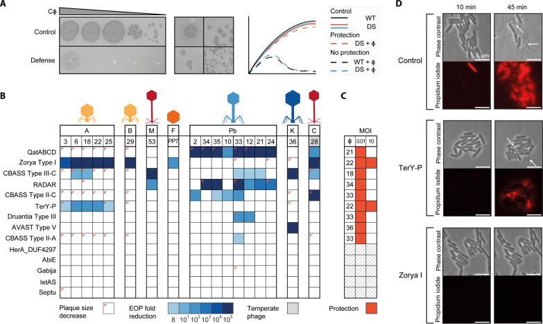
Fig. 3. Defense systems provide genera-specific anti-phage activity.
(A) Representation of the assays used to assess the effect of individual DS on phage infectivity. Left: An example of fold decrease in ϕPa33 phage infectivity caused by the defense system QatABCD. Middle: Examples of phage plaque size decrease observed for Autographiviridae ϕPa3 and Casadabanvirus ϕPa28 with QatABCD cells. Right: Illustrations of growth curves obtained from liquid culture collapse assays for control strains (wild type, WT) and strains containing individual DS with and without phage infection, showing cases of protection (orange) and no protection (blue). (B) Efficiency of plating (EOP) of phages in PAO1 containing individual DS. EOP was determined as the fold decrease of phage titer in the strain with the defense system compared to the titer obtained in the strain without the defense system. Plaque size reductions are indicated as a colored corner. (C) Liquid culture collapse assays of PAO1 containing individual DS infected with phages, as compared to a control (WT) without a defense system. Results are shown as a summary of the effects observed when infecting the cultures with a multiplicity of infection (MOI) of 10 or 0.01. A protective effect is represented as dark orange. The absorbance values at 600 nm of representative phage-host combinations are shown in fig. S8. (D) Time-lapse phase contrast and fluorescence images of PAO1 cells containing individual DS infected with phage ϕPa25. Cells were stained with propidium iodide to visualize the permeabilization of the cell membrane due to cell death. TerY-P and Zorya Type I cells survive phage infection, although some cell death is still observed for TerY-P, consistent with the EOP results in (B).

