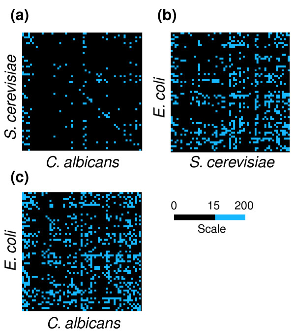
Figure 8.

Differential display maps for comparative analysis of codon context. To compare the codon context maps of different species, the order of the codons displayed in the map was fixed and the maps overlapped using a differential display tool built into the Anaconda bioinformation system. Maps representing the context differences between (a) S. cerevisiae and C. albicans, (b) E. coli and S. cerevisiae and (c) C. albicans and S. cerevisiae were obtained by calculating the module of the difference between the residuals of each map. The differences are represented in blue according to the color scale. The blue cells indicate the highest context difference and the black cells represent pairs of codons that have similar residual values between two species (module of the difference between residuals falls within the 0-15 interval). The maps show rather large differences in codon context between E. coli and S. cerevisiae or C. albicans and smaller differences between S. cerevisiae and C. albicans.
