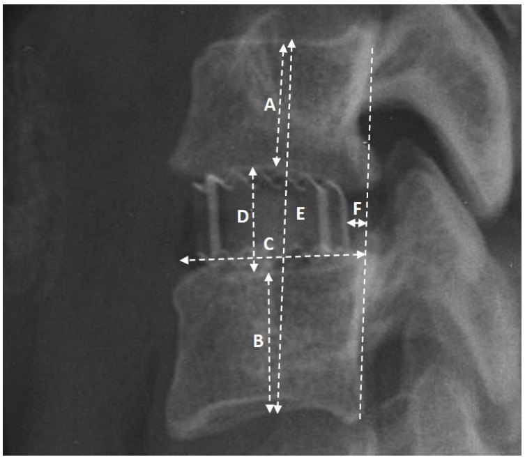
Figure 2.
The image of the operated segment in the lateral projection, showcasing the selected radiological parameters utilized in the study: A—height of the upper body; B—height of the lower body; C—length of the lower endplate; D—height of the intervertebral space; E—height of the segment; F—distance of the implant from the medial column (posterior longitudinal ligament).

