Figure 1.
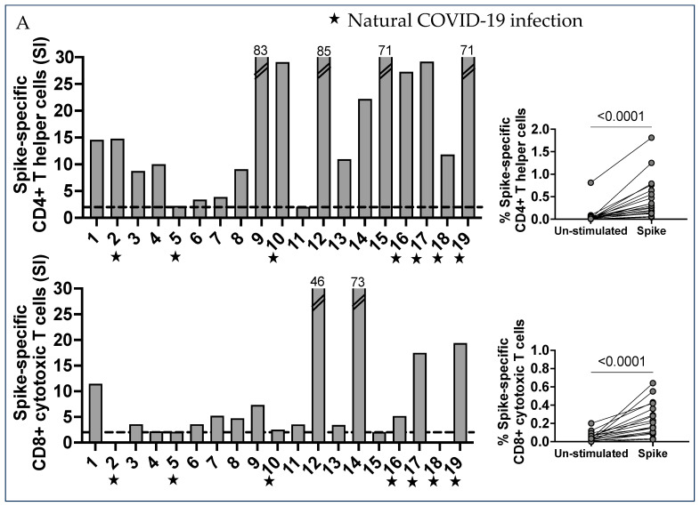
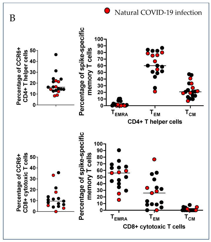
Characterization of SARS-CoV-2 spike-specific CD4+ and CD8+ T cells and the development of T cell memory. (A) PBMC were separated from 19 vaccinated pregnant women who received 3 injections of mRNA-based vaccination for COVID-19 protection. PBMC were stimulated in vitro with the spike peptides pool, which includes 253 peptides that are 15 amino acids long, overlapping by 10 amino acids, and spanning the entire spike protein. Then, 24 h after stimulation, the cells were collected and stained with specific monoclonal antibodies to determine the T cell activation state in response to peptide antigens (AIM assay). The percentage of AIM+ CD4+ Th cells from the un-stimulated control and SARS-CoV-2 CD4 spike-stimulated cells, presented as stimulation index (SI) values, were compared using the Wilcoxon matched-pairs signed rank test. The percentage AIM+ T cell increase for the unstimulated samples was a positive T cell response: a stimulation index of 2 (dotted lines) was considered a good T cell response. The percentage of AIM+ CD8+ CTL cells from un-stimulated control and SARS-CoV-2 CD4 S-all-stimulated cells is also presented here, using the stimulation index (SI). Results were compared by the Wilcoxon matched-pairs signed rank test. CCR6 expression was found in a large percentage of SARS-CoV-2 spike-specific CD4+ Th cells in most subjects, and CD8+ CTL cells were also expressing CCR6. (B) T cell memory phenotypes. Memory populations were defined by specific markers on gated AIM+ CD4+ Th and CD8+ CTL cells. Each dot shows the percentage of memory populations: terminally differentiated effector T cells (CD45RA+ CCR7- TEMRA), effector memory T cells (CD45RA- CCR7- TEM), and central memory T cells (CD45RA- CCR7+ TCM).


