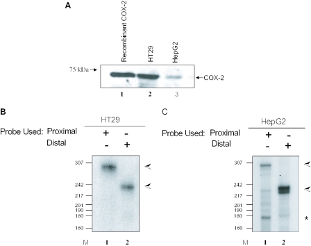
Figure 2.
Tissue-specific alternative polyadenylation of COX-2 in HT29 and HepG2 cells. (A) Western blot of 75 μg total protein cell extract from HT29 and HepG2 cells using an anti-COX-2 monoclonal antibody. Lane 1, 1 μg recombinant COX-2 protein. (B and C) RNase protection assays using endogenous RNA from HT29 (B) and HepG2 (C) cells along with COX-2 proximal and/or distal probes. The data shown in (B), lane 2, illustrate that HT29 cells primarily use the COX-2 distal polyadenylation signal. (C) HepG2 cells utilizing the COX-2 proximal signal in lane 1 and the distal signal in lane 2. The band corresponding to utilization of the COX-2 proximal polyadenylation signal is marked with an asterisk; the band corresponding to utilization of the COX-2 distal polyadenylation signal is marked with an arrow.

