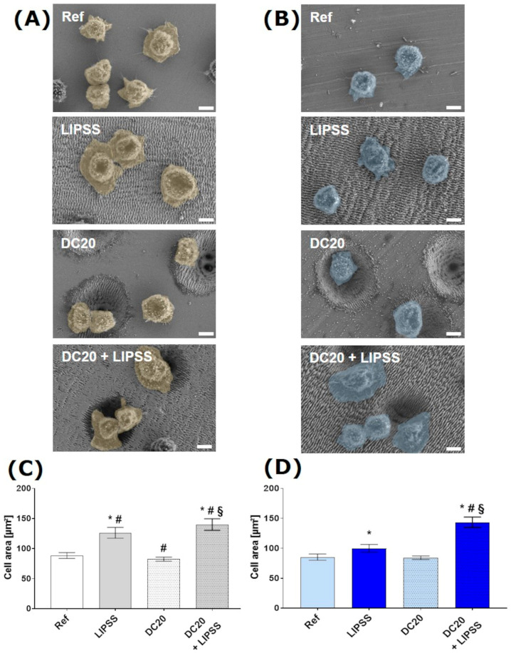
Figure 4.
Cell spreading and growth of human keratinocytes (HaCaT) after 2 h on laser-structured metal specimens: (A) on medical steel and (B) on titanium alloy (Ti6Al4V). (FE-SEM Merlin VP compact, 2000× magnification, 5 kV, HE-SE detector, scale bars = 5 µm). The cell area of HaCaTs on (C) steel and (D) on Ti6Al4V. Note that the spreading of cells on both metals with micro-dimple clusters and additional nanostructures (DC20 + LIPSS) is significantly increased compared to all other specimens. Also, cells on the laser-induced periodic surface structures (LIPSS) shown on both materials achieved an enhanced cell area compared to polished metals (Ref). (ImageJ; n = 40 cells of SEM images, mean ± s.e.m., Kruskal–Wallis test post-tested with adjusted Mann–Whitney test: * p ≤ 0.05 to Ref, # p ≤ 0.05 to LIPSS, § p ≤ 0.05 to DC20).

