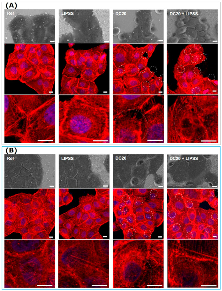
Figure 6.
Cell morphology and actin cytoskeleton organization of human keratinocytes (HaCaT) after 24 h on different laser-structured metal specimens: (A) medical steel and (B) Ti6Al4V. Note that the cells grew on both metals and were independent of the structuring process. After 24 h, there was no apparent change in cell morphology and actin cytoskeleton. HaCaTs could span over the micro-dimple clusters and their actin filaments. (First row: FE-SEM Merlin VP compact, 1000× magnification, 5 kV, HE-SE detector. Second and third rows: LSM780, 63× oil objective, both Carl Zeiss; actin in red, nucleus in blue, underlying micro-dimples are marked as dotted circles, scale bars = 10 µm).

