Figure A1.
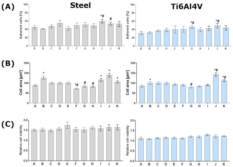
Cellular responses of human keratinocytes (HaCaT) on different specimens within 24 h: (left) on medical steel and (right) on titanium alloy (Ti6Al4V). Note that specimens A-Ref, B-LIPSS, H-DC20, and J-DC20 + LIPSS, respectively, were presented in the article. (A) Results regarding initial adhesion after 10 min show that only J (DC20 + LIPSS) could increase cellular attachment on both materials (FACSCalibur; n = 4, mean ± s.e.m.). (B) The spreading capacity of HaCaTs on structure J (DC20 + LIPSS) was most pronounced. The cell area was significantly improved on nanostructure B (LIPSS) and another nano- and microtopography combination (K). However, these structures did not show a significant increase in initial adhesion. It should be noted here that laser structures both promote and inhibit cell spreading (ImageJ; n = 40 cells of SEM images, mean ± s.e.m.). (C) Viability data show no effects on metabolic activity. However, the lower viability of HaCaT on Ti6Al4V than medical steel should be noted (Anthos reader; n = 4, mean ± s.e.m). (Statistics: adjusted Mann–Whitney test: * p ≤ 0.05 to Ref, # p ≤ 0.05 to LIPSS.)

