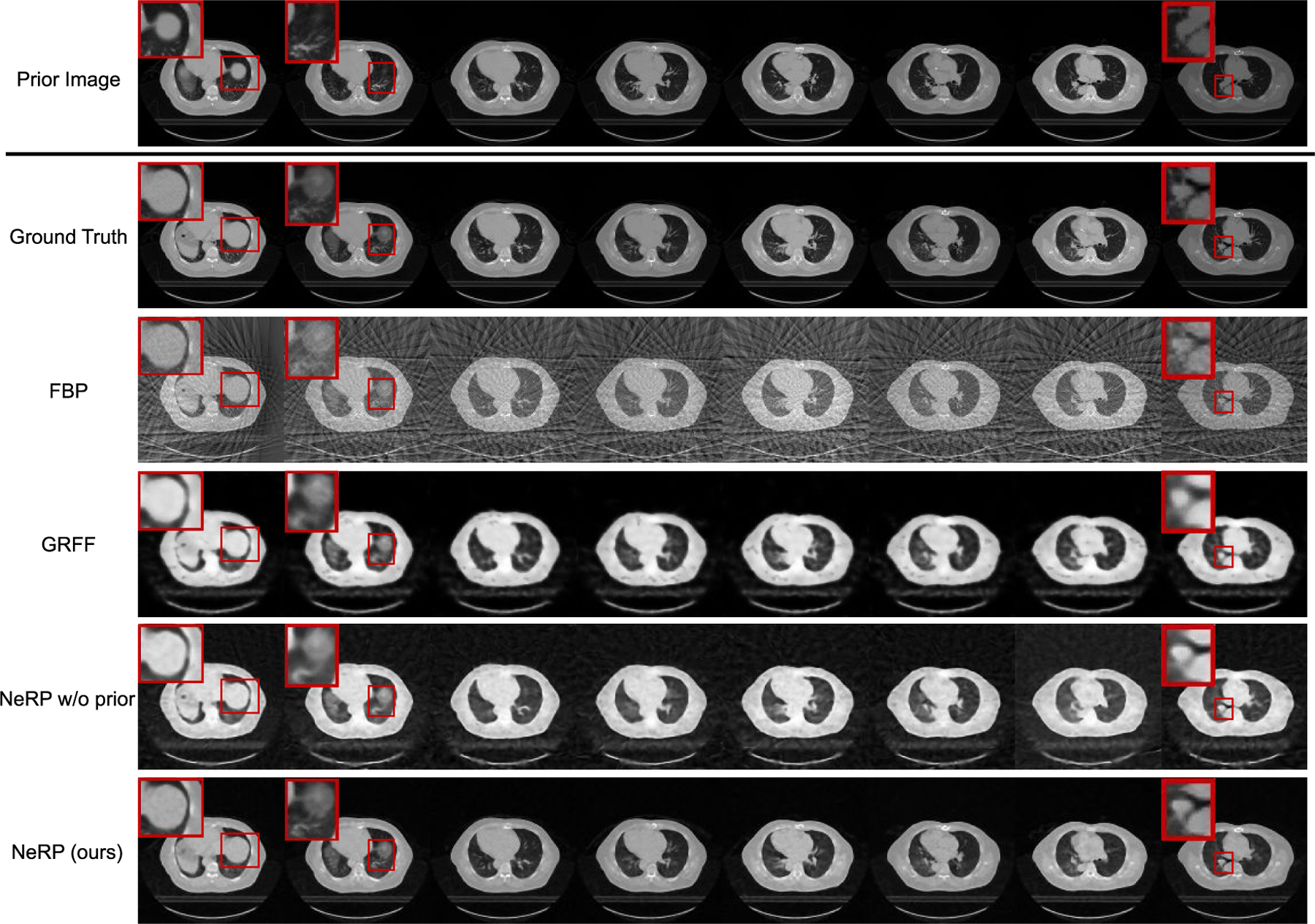
Fig. 3.

Results of 3D CT image reconstruction for pancreas 4D CT data using 20 projections. The first and second rows show the prior 3D CT (phase 1) and the ground truth of target 3D CT (phase 6) image, where each column demonstrates cross-sectional slices of the 3D volume. The final row shows the reconstructed 3D CT images by using the proposed NeRP method. For comparison, the second to fourth rows show the reconstruction results of FBP method, GRFF method [20] and NeRP without using prior embedding. The zoom-in red boxes highlight the difference in anatomic structure for comparison.
