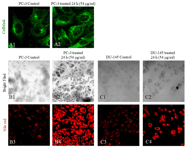
Figure 6.
Cell morphology imaging using CellMask™ and Nile red. Following incubation with 54 μg/mL FALS for 24 h, cells grown on glass coverslips were stained and visualized under a confocal microscope. (A1) Untreated PC-3 cells (magnification ×630) and (A2) treated PC-3 (magnification ×630) did not exhibit any visible membrane deformities following treatment with FALS; (B1) bright-field image of untreated PC-3 cells (magnification ×400) and (B2) image after incubation with FALS (magnification ×400); (B3) staining of untreated PC-3 cells with Nile red. The excitation was performed at 488 nm, and the emission at 565 nm was captured. The dim fluorescent intensity is due to the staining of intracellular lipids in the endoplasmic reticulum (ER); (B4) treated PC-3 cells exhibit bright fluorescence because of intracellular lipid accumulation in the ER (magnification ×400). (C1) Regarding cell morphology, analogous observations were made in the DU-145 cell line (magnification ×400), where granulation was amplified in the treated cells (magnification ×400) (C2); (C3) the untreated DU-145 cells exhibited dim fluorescence in basal conditions (magnification ×400), while; (C4) the treated cells had granulous stained areas, which are thought to be lipid droplets inside the ER (magnification ×400).

