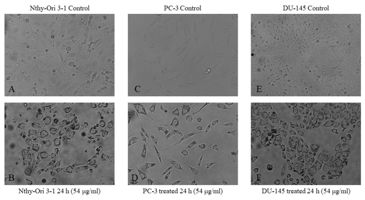
Figure A5.
Cell morphology under a phase-contrast microscope. Following incubation with 54 μg/mL FALS for 24 h, cells grown on glass coverslips were visualized under a phase contrast microscope. All photographs were taken at ×200 magnification. (A) Untreated Nthy-Ori 3-1 cells; (B) treated Nthy-Ori cells where granulation and darker colors are visible; (C) untreated PC-3 cells; (D) treated PC-3 cells; (E) untreated DU-145 cells; and (F) treated DU-145 cells. All cell lines exhibited darker coloration, increased and abnormal size, and visible granulation.

