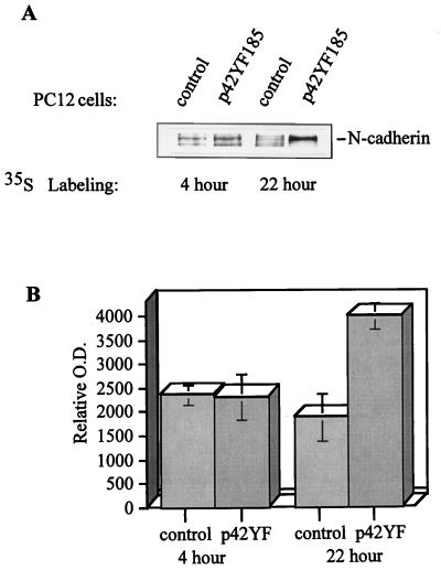
FIG. 9.
Metabolic labeling demonstrates that N-cadherin synthesis is increased in p42YF185 cells compared to that in mock-transfected cells. (A) N-cadherin was immunoprecipitated from cell lysates, separated by SDS-PAGE, and visualized by autoradiography. (B) The relative amounts of newly synthesized N-cadherin in control versus p42YF185 cells were quantified. O.D., optical density.

